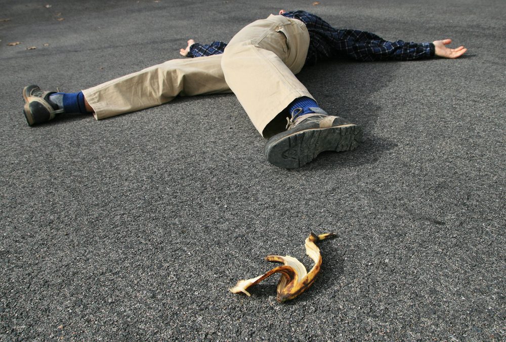
A young healthy person slips on a banana peel and inverts his ankle.
X-rays reveal:
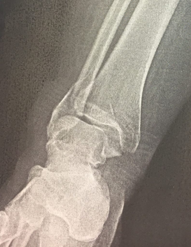
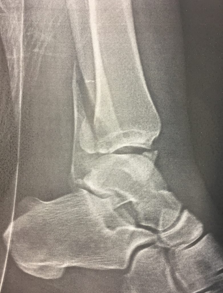
This is a trimalleolar fracture. Technically a person only has two malleoli (medial and lateral). A trimalleolar fracture involves both of these along with the posterior aspect of the tibial plafond (referred to as the posterior malleolus). Having three parts, it is a more unstable fracture and may be associated with ligamentous injury.
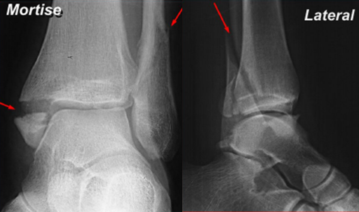
In fact, you may only find a lateral and posterior malleolar fracture on x-ray, with a widened ankle mortise. This may represent a tear of the deltoid ligament and is considered a ‘trimalleolar equivalent’.

In the above x-ray, for instance, you can see the bimalleolar fracture along with a widened mortise. This is a ‘trimalleolar equivalent’.
ED management is to provide a ‘cadillac’ splint (posterior and U-shaped), crutches, ice, and non-weight-bearing status, with close orthopedics follow-up. These almost universally require operative intervention. If there are other injuries or the patient appears unreliable or overtly uncomfortable with the plan, consider admission.
In some instances, patients may required sedation and closed reduction prior to discharge.

